
Figure 3.
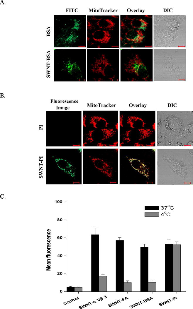
Subcellular localization of surface-modified SWNTs. A. Subcellular localization of BSA-FITC and SWNT-PL-PEG-BSA-FITC in HeLa cells. Cells transfected with CFP-lamp and stained with MitoTracker were incubated with BSA-FITC (1 μg/ml of BSA) or SWNT-PL-PEG-BSA-FITC (2.5 μg/ml of SWNTs) for 30 minutes. Confocal images of the cells show that BSA-FITC and SWNT-PL-PEG-BSA-FITC are mainly localized in the lysosome. B. Subcellular localization of PI and SWNT-PL-PEG-PI in HeLa cells. Cells transfected with CFP-lamp and stained with MitoTracker were incubated with PI (5 μg/ml) or SWNT-PL-PEG-PI (2.5 μg/ml of SWNTs) for 30 minutes. Confocal images of the cells show that SWNT-PL-PEG-PI is mainly localized in the mitochondria. C. Statistical analysis of fluorescence emission intensity of surface-modified SWNTs in cells incubated at 37°C or 4°C. Only small, non surface-targeting molecules (PI) can be carried by SWNTs into the mitochondria without being affected by the loss of cell endocytosis function. Bars, means SD (n=5).