
Figure 4.
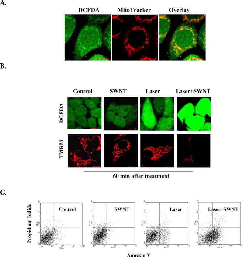
Apoptosis induced by mitochondrial SWNTs under laser irradiation. HeLa cells were incubated with SWNT (2.5 μg/ml) for 2 hours, then irradiated with a 980 nm laser (0.75 W/cm2) for 2 minutes. A. Initial ROS generation in the mitochondria after laser irradiation. B. ROS generation and ΔΨm changes in the cells at different time points after treatment. Fluorescent image series of H2DCFDA and TMRM were acquired after different treatments. C. Cell apoptosis induced by different treatments. Cells harvested 6 hours after different treatments were double stained by Annexin V and PI, and analyzed by FACS.