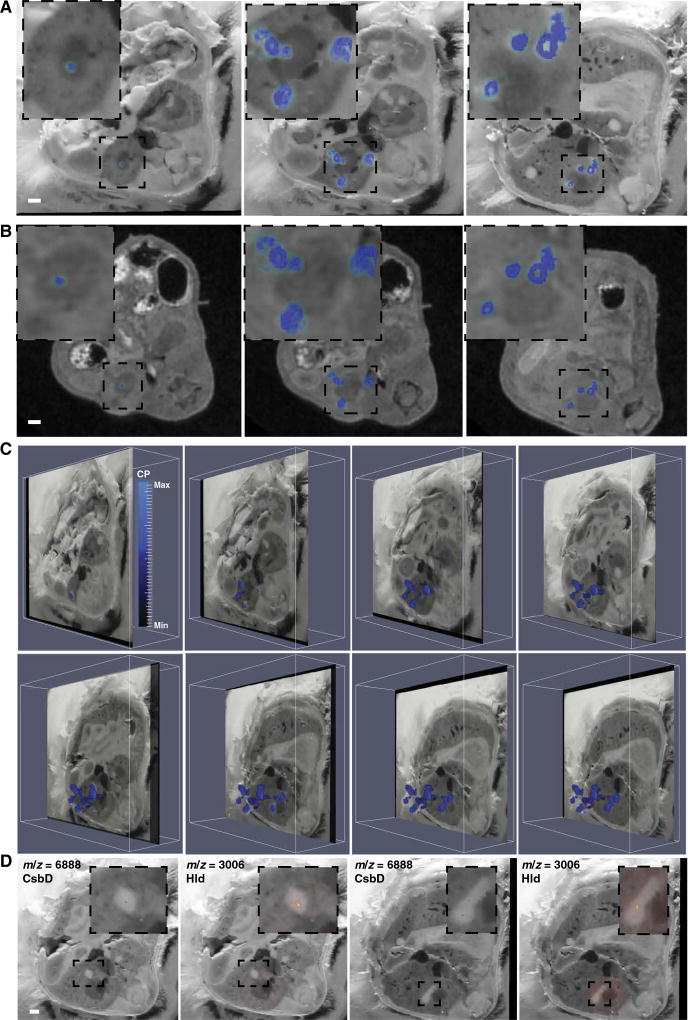
Fig. 4. MS imaging delineates bacterial and host proteins at the infectious interface.
MALDI IMS performed on serial transverse sections of the region of tissue encompassing the right kidney in a mouse infected with S. aureus 4 days before. A spectral peak at m/z = 10,164, subsequently identified as the S100A8 subunit of the heterodimeric protein calprotectin, colocalized with abscesses when MALDI IMS data were co-registered to blockface imaging (A) or MRI (B). Three representative sections are shown. Insets: Enlarged image showing only the right kidney. White bars represent 1-mm scale. (C) Obliquely oriented serial transverse blockface images with overlaid MALDI IMS data to depict the 3D orientation of S100A8 (m/z = 10,164). Heat map depicts minimum and maximum threshold values in arbitrary units. (D) Blockface images overlaid with MALDI IMS for m/z = 3006 (subsequently identified as δ-hemolysin) and m/z = 6888 (subsequently identified as CsbD-like protein), which are bacterial proteins colocalizing to the nidus within an abscess. Two representative blockface sections are shown. White bar represents 1-mm scale. Insets: Enlarged images showing only the abscess and immediately surrounding tissue. See also movies S6 to S8.

