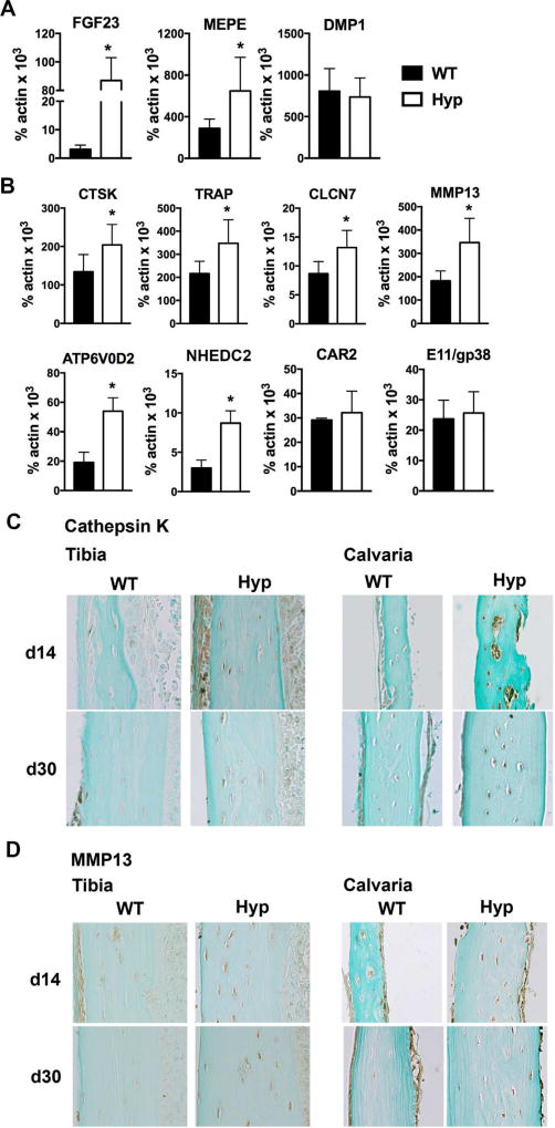
Fig. 3.
Hyp osteocytes have increased expression markers of perilacunar and canalicular remodeling. (A) RNA was isolated from osteocyte enriched WT and Hyp femoral cortices and subjected to RT-qPCR to quantitate mRNA expression of FGF23, MEPE, and DMP1. (B) mRNA expression of genes implicated in osteocyte perilacunar/canalicular remodeling. (C) Immunohistochemistry for cathepsin K on d14 and d30 tibia and calvaria. (D) Immunohistochemistry for MMP13 on d14 and d30 tibia and calvaria. Data are representative of that obtained from 3 mice per genotype/age. *p < 0.05.

