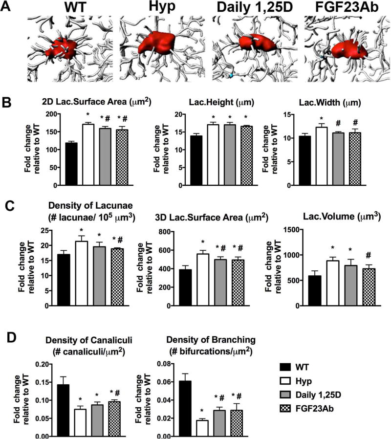
Fig. 5.
Treatment of Hyp mice with 1,25D or FGF23Ab partially restores calvarial lacunocanalicular network morphology. Intravital THG microscopy was performed on d30 mice. (A) Imaris software was used to produce 3D images of the osteocyte lacunae and canaliculi. (B) Evaluation of 2D lacunar (lac.) surface area, width, and height. (C) Quantitation of lacunar density, 3D surface area, and volume. (D) Quantitation of canalicular density and density of branched canaliculi extending from each lacuna. Data are representative of that obtained from 4 mice per genotype/treatment group. *p < 0.05, #p < 0.05 versus Hyp.

