Figure 3.

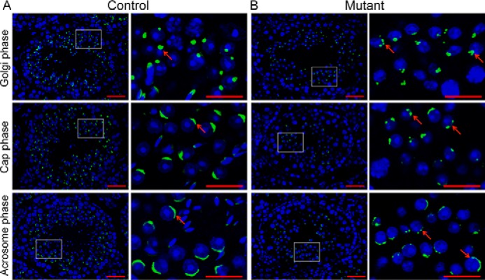
Abnormal acrosome biogenesis in Setd2 cKO mouse spermatids. Shown is histochemical staining of FITC-conjugated PNA (green) and DAPI (blue) in sections of adult control (A) and Setd2 cKO (B) mouse testes. The Golgi, cap, and acrosome phases of spermiogenesis are shown in order. A, PNA staining of representative testes from adult controls. Acrosomes are labeled by PNA (green). The arrow indicates a representative acrosome. In the Golgi phase, the acrosome displays a single granule close to the nucleus. In the cap phase, the acrosome grows to form the cap structure covering the nucleus. In the acrosome phase, the acrosome forms the moon-shaped structure covering the nucleus. B, PNA staining of representative testes from adult Setd2 cKO mice. There are multiple PNA-positive structures (as indicated by the arrows) in Setd2 cKO mouse spermatids throughout spermiogenesis. Right panels show higher-magnification views of the framed areas in the left panels (scale bars, 40 μm).
