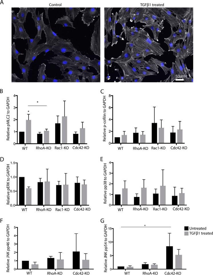
Figure 5.
Cdc42-KO MSC display increased pJNK. A, F-actin of TGFβ1-treated and untreated MSC detected by fluorescently labeled phalloidin. B–G, quantifications of Western blot analyses of lysates of indicated MSC for pMLC2 (B), pCofilin (C), pERK (D), pp38 (E) JNK pp46 (F), and JNK pp54 (G). (black bars, 24-h TGFβ; gray bars, untreated; n = 3/3).

