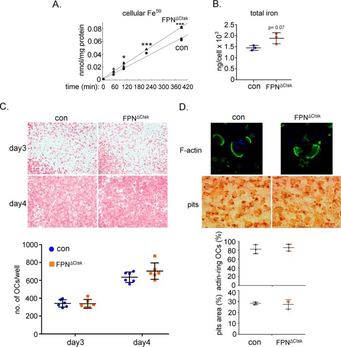
Figure 4.
Deletion of Fpn in mature osteoclasts has no effects on osteoclast differentiation and function. Bone marrow monocytes were cultured with M-CSF (BMM) or M-CSF + RANKL for 2 and 4 days to generate mononuclear pre-osteoclasts (pOC) and mature multinucleated osteoclasts (OC), respectively. A, intracellular levels of uptaken Tf-Fe59 in mature osteoclasts were measured by a gamma counter. The data were normalized by protein concentrations. n = 3. B, amount of cellular total iron in mature osteoclasts was measured using a colorimetric iron assay method. The data were normalized by osteoclast number in each well of a 6-well plate. n = 3. C, TRAcP staining of osteoclasts cultured on 48-well plastic tissue culture plates. The number of TRAcP+ osteoclasts with more than three nuclei was counted. n = 6. D, confocal microscopic images (upper panels) of filament actin (F-actin, green) and nucleus (blue) staining in osteoclasts cultured on bone slices. Resorption pits (lower panels) were stained with HRP-labeled WGA lectin. The percentage of active osteoclasts (OCs with actin rings) to the total number of osteoclasts with more than three nuclei was quantified. The pits area (%) was measured and calculated using ImageJ software (National Institutes of Health). The data shown are representatives from three independent experiments. *, p < 0.05; **, p < 0.01; ***, p < 0.001 versus control (con) by one-way ANOVA.

