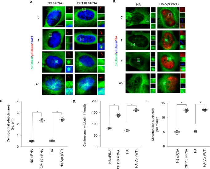
Figure 10.
Depletion of CP110 or expression of Vpr enhances microtubule nucleation. A, HeLa cells transfected with nonspecific (NS) or CP110 siRNA were subjected to a microtubule regrowth assay. Cells were processed for immunofluorescence at the indicated time points after release and stained with antibodies against α-tubulin (green) and γ-tubulin (red). DNA was stained with DAPI (blue). B, HeLa cells transfected with plasmid expressing HA or HA-Vpr(WT) were subjected to a microtubule regrowth assay. Cells were processed for immunofluorescence at the indicated time points after release and stained with antibodies against HA (red), α-tubulin (green), and γ-tubulin (blue). C, the staining area of γ-tubulin at the centrosome was quantitated. D, the staining intensity of γ-tubulin at the centrosome was quantitated. E, the number of cytoplasmic microtubules emanated from the centrosome was determined at the 1-min time point. For C–E, at least 20 cells were scored for each condition in each experiment, and the mean (thick open line) and standard error (bar) of three independent experiments (○) are shown in the graph. *, p < 0.01.

