Figure 6.
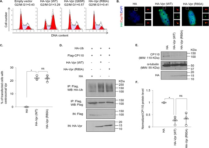
Vpr centrosomal localization and Vpr-induced ubiquitination and degradation of CP110 are independent of G2/M arrest. A, HEK293T cells were co-transfected with plasmids expressing GFP and HA (Empty vector), HA-Vpr(WT), HA-Vpr(Q65R), or HA-Vpr(R80A). Cell cycle profiles were determined by flow cytometry gating on the GFP+ population. The G2/M:G1 ratio is presented for each condition. B, HeLa cells transfected with plasmid expressing HA, HA-Vpr(WT), or HA-Vpr(R80A) were processed for immunofluorescence and stained with antibodies against HA (green) and Cep78 (red). DNA was stained with DAPI (blue). Scale bar, 2 μm. C, the percentage of HA-expressing cells showing centrosomal localization of Vpr was determined. At least 100 cells were scored for each condition in each experiment, and the mean (thick open line) and standard error (bar) of three independent experiments (○) are shown in the graph. *, p < 0.01; ns, nonsignificant. D, HEK293 cells were co-transfected with plasmids expressing HA-Ub, FLAG-CP110, and HA, HA-Vpr(WT), or HA-Vpr(R80A). Lysates were immunoprecipitated (IP) with an anti-FLAG antibody in 1% SDS and Western-blotted (WB) with the indicated antibodies. IN, input. E, HEK293 cells were transfected with plasmid expressing HA, HA-Vpr(WT), or HA-Vpr(R80A). Lysates were Western-blotted with the indicated antibodies. α-Tubulin was used as loading control. F, normalized CP110 protein level. The mean (thick open line) and standard error (bar) of three independent experiments (○) are shown in the graph. *, p < 0.01.

