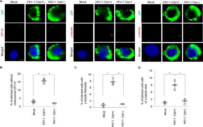
Figure 9.
HIV-1 Vpr induces CP110 loss, centriole elongation, and centrosome amplification in infected T-cells. A, MT4 cells, mock-infected or infected with either WT HIV-1 (Vpr+) or HIV-1 missing Vpr (Vpr−), were processed for immunofluorescence and stained with antibodies against p24 (green) and CP110 or γ-tubulin (red). DNA was stained with DAPI (blue). Scale bar, 2 μm. B, the percentage of p24-positive cells with no centrosomal CP110 staining was determined. C and D, the percentage of p24-positive cells with elongated centrioles (γ-tubulin filaments) (C) or centrosome amplification (>2 γ-tubulin dots) (D) was determined. For B–D, at least 100 cells were scored for each condition in each experiment, and the mean (thick open line) and standard error (bar) of three independent experiments (○) are shown in the graph. *, p < 0.01.

