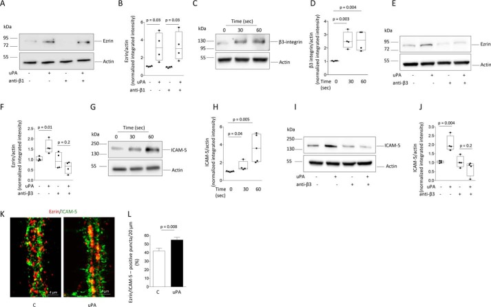
Figure 3.
β3-Integrin and ICAM-5 mediate uPA-induced recruitment of ezrin to the postsynaptic density. A and B, representative immunoblot (A) and mean intensity of the band (B) of Ezrin expression in PSD extracts prepared from WT cerebral cortical neurons incubated 0–60 s with 5 nm of uPA, alone or in the presence of 10 μg/ml of anti–β1-integrin blocking antibodies. The data are expressed as means ± S.E. for n = 4 observations. C and D, representative immunoblot (C) and mean intensity of the band (D) of β3-integrin expression in PSD extracts prepared from WT cerebral cortical neurons incubated 0–60 s with 5 nm of uPA. The lines denote S.E. The data are expressed as means ± S.E. for n = 4 observations. E and F, representative immunoblot (E) and mean intensity of the band (F) of ezrin expression in PSD extracts prepared from WT cerebral cortical neurons treated during 60 s with 5 nm of uPA, alone or in the presence of 10 μg/ml of anti–β3-integrin blocking antibodies. The data are expressed as mean ± S.E. for n = 4 observations. G–J, representative immunoblot (G and I) and mean intensity of the band (H and J) of ICAM-5 expression in PSD extracts prepared from WT cerebral cortical neurons incubated 0–60 s with 5 nm of uPA, alone (G and H) or in the presence of 10 μg/ml of anti–β3-integrin blocking antibodies (I and J). The data are expressed as means ± S.E. for n = 4 observations. K, representative confocal microscopy pictures taken at 40× magnification of dendrites from WT cerebral cortical neurons stained with anti-ezrin (red) and anti-ICAM-5 (green) antibodies following 60 s of incubation with 5 nm of uPA or vehicle (control: C). L, mean percentage of ezrin-positive puncta that co-localizes with ICAM-5 in dendrites of 40 WT neurons from three different cultures incubated with uPA or vehicle (control) as described for I. The data are expressed as means ± S.E. for n = 30 observations per experimental group.

