Figure 6.
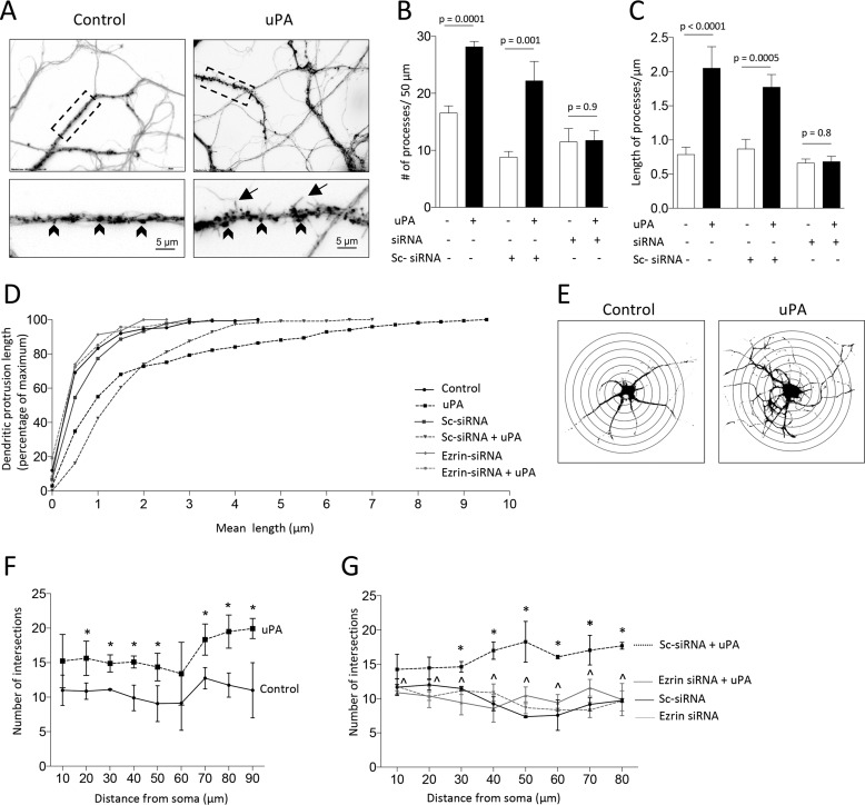
Ezrin mediates uPA-induced formation of dendritic protrusions and branches. A–C, representative micrographs (A) and mean number (B) and length (C) of dendritic protrusions/50 μm in 300 WT cerebral cortical neurons from three different cultures treated for 0–60 min with 5 nm of uPA or a comparable volume of vehicle (control), alone or in the presence of ezrin siRNA or scramble (Sc) siRNA. Magnification in A, 20× in upper panels. Lower panels correspond to a 60× magnification of the area denoted by the dashed rectangle in each upper panel. The lines denote S.E. Arrowheads denote dendritic spines; arrows depict filopodia. D, cumulative frequency plot depicting the mean length of dendritic protrusions in the 300 neurons examined in A–C. E–G, representative Sholl analysis (E) and mean number of dendritic intersections (F and G) in WT cerebral cortical neurons treated 60 min with 5 nm of uPA, alone (F) or in the presence of either ezrin siRNA or Sc-siRNA (G). n = 400 neurons in F and 300 neurons in G. Asterisks in F denote p < 0.05 compared with number of intersections at a comparable distance from the soma in control neurons. Asterisks in G denote p < 0.05 when cells treated with Sc-siRNA and uPA are compared with cells incubated with Sc-siRNA and vehicle (control). *, p < 0.05 when cells treated with ezrin siRNA and uPA are compared with neurons treated with ezrin siRNA and vehicle (control). The lines denote S.E.

