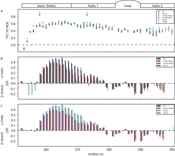
Figure 3.
A–C, NMR derived picosecond–nanosecond timescale dynamics (A) and secondary structures (B and C) of Myc bHLH and its variants, including the schematics of Myc bHLH-LZ secondary structure. A, heteronuclear 15N-1H NOE of Myc bHLH-LZ and its variants. pThr-358 and pSer-373 phosphorylation sites are marked with arrows. B and C, secondary structure propensities of Myc bHLH and variants obtained from Cα and Cβ chemical shifts.

