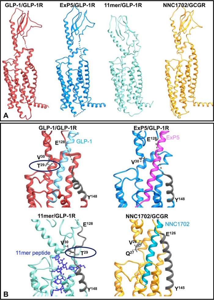
Figure 7.
Peptide-bound full-length structures of GLP-1R and GCGR. A, full-length structures illustrating the relative position of the N-terminal ECD to the receptor core. B, zoom-in of the resolved far N-terminal residue(s) and TM1/ECD stalk (highlighted in dark gray). The backbones of the peptide agonists are illustrated in ribbon (GLP-1, exendin-P5 (ExP5), and NNC1702) or X-stick (11-mer).

