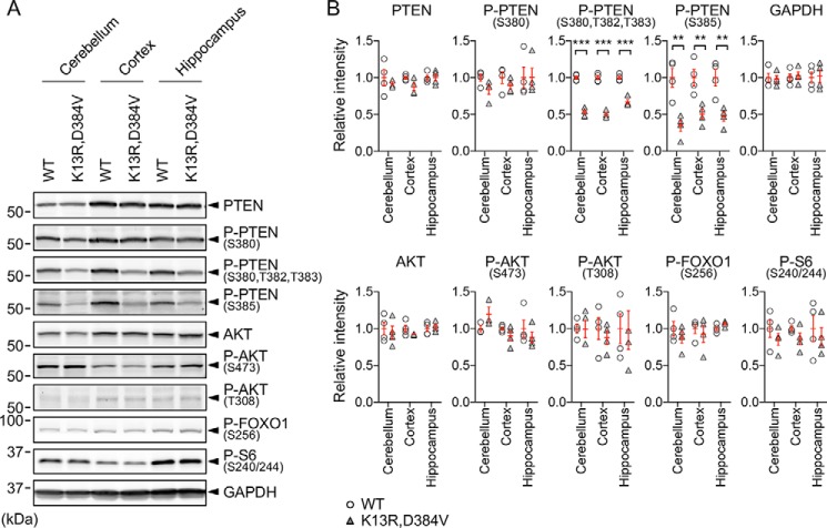
Figure 7.
PI3K signaling was not affected in PTENK13R, D384V mice. A, Western blotting of the cerebellum, cortex, and hippocampus isolated from 3-month-old PTENK13R, D384V and littermate control mice using antibodies to PTEN, phospho-PTEN (S380), phospho-PTEN (S380, T382, T383), phospho-PTEN (S385), AKT, phospho-AKT (S473), phospho-AKT (T308), phospho-FOXO1 (S256), phospho-S6 (S240/244), and GAPDH. B, quantification of band intensity. Bars are the mean ± S.E. (n = 4). Statistical analysis was performed using the Student's t test: **, p < 0.01; ***, p < 0.001.

