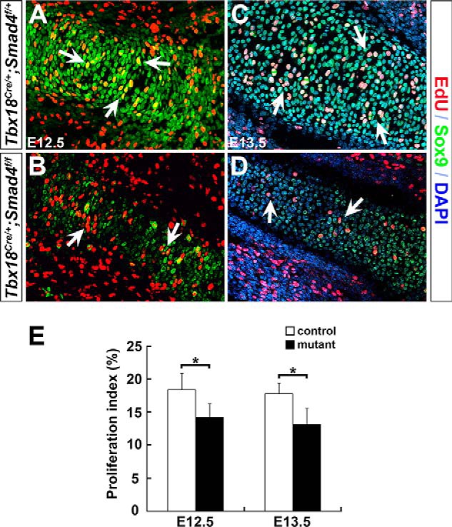
Figure 4.

Decreased chondrocyte proliferation in Smad4 mutant mice. A–D, chondrocyte proliferation was analyzed by EdU labeling (red). Sox9 (green) was used to mark chondrocytes. Arrows indicate EdU-positive chondrocytes. E, EdU-positive cells were quantified. The proliferation rate of chondrocytes was represented by the ratio of EdU/Sox9 double positive cells normalized to Sox9 positive cells in humerus. n = 3, *, p < 0.05.
