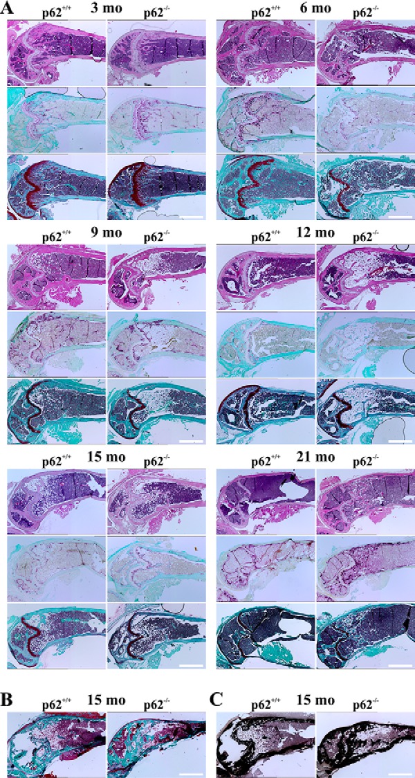
Figure 8.

A, representative examples of microscopy images of H&E- (top), TRAP- (middle), and Safranin O (bottom)-stained paraffin sections of decalcified femora with WT or p62−/− origin at 3, 6, 9, 12, 15, or 21 months (mo) (C57BL/6N). Note the age-dependent lipid accumulation in p62-deficient bones. Tb.N and TRAP activity are elevated, and Tb.Sp is reduced in p62−/−-derived bones, especially at 15 and 21 months. B and C, microscopy images presenting sections of MMA-embedded nondecalcified bones (C57BL/6N) after Masson-Goldner trichrome (B) or von Kossa staining (C). Scale bars, 1 mm.
