Figure 9.

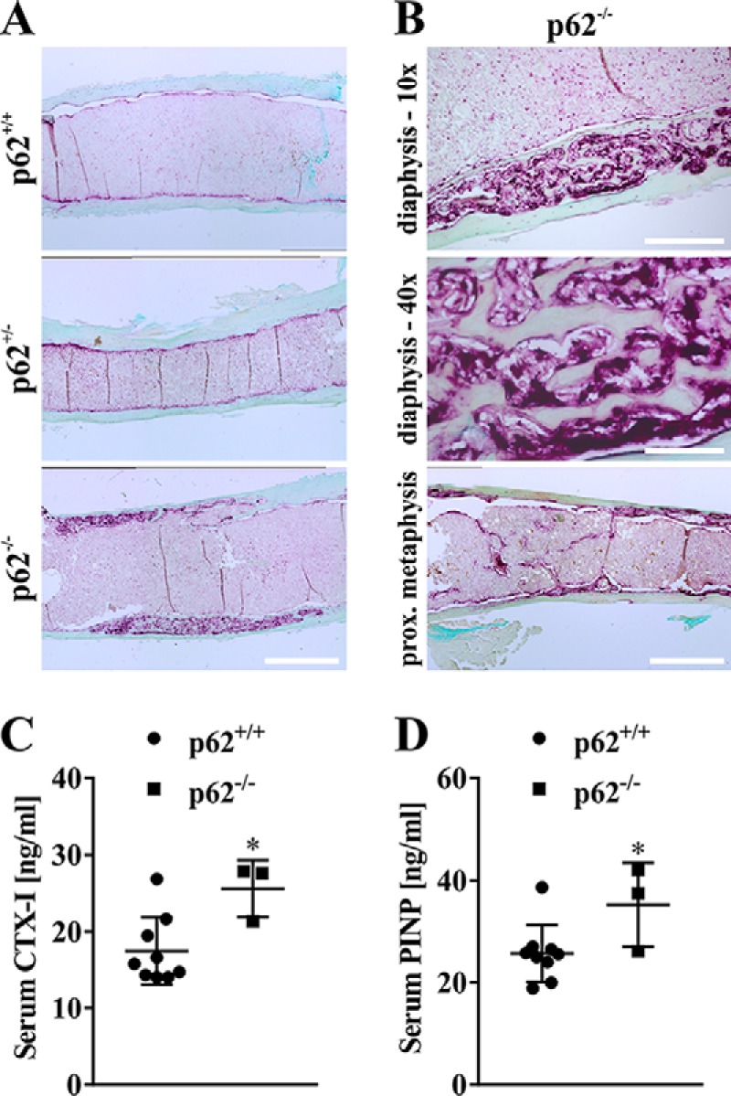
A, representative microscopy images of TRAP-stained bone sections from female p62+/+, p62+/−, and p62−/− mice at 21 months indicate PDB-like osteolytic lesions in femora (distal diaphysis) of p62-deficient animals (C57BL/6N). Scale bars, 1 mm. B, osteolytic lesions and TRAP activity in diaphysis of p62−/−-derived bones from A are shown at higher magnification (scale bars, 300 μm (upper image) and 75 μm (middle image)). Increased trabecular material combined with pronounced TRAP activity exclusively detected in proximal metaphysis of p62 KO female animals is exemplified in the lower image (scale bar, 1 mm). C and D, quantification of bone degradation marker CTX-I (C) and bone formation marker PINP (D) in serum of p62+/+ and p62−/− mice (C57BL/6N). Data are displayed as the mean ± S.D. (error bars). Significance is indicated by asterisks (*, p < 0.05, Student's t test).
