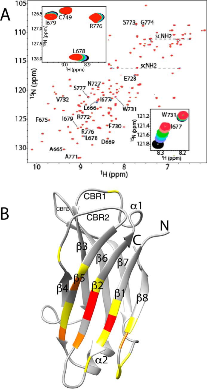
Figure 4.

C2(C771A)–PI(3)P interaction monitored by NMR. A, overlay of C2(C771A) 1H-15N HSQC spectra as a function of increasing PI(3)P concentration. Perturbed backbone NH peaks are labeled, and the two perturbed but unassigned side-chain NH2 peak pairs are connected by dashed lines and denoted as “scNH2.” The insets highlight the chemical shift changes of backbone NH peaks of Ile-677 (strand β2) and Trp-731 (β5) and of Leu-678 (β2), Ile-679 (β2), and Arg-776 (β7-β8 loop). Peak colors and corresponding C2(C771A):PI(3)P ratios are as follows: red, 1:0; pink, 1:0.25; green, 1:0.5; cyan, 1:0.75; purple, 1:1; black, 1:1.5. B, perturbed residues mapped onto a ribbon representation of C2(C771A). Yellow represents residues with Δδav (ppm) ≤ 0.01, orange represents 0.01–0.03, and red represents >0.03 where Δδav (ppm) = ((ΔδH2 + ΔδN2(γN/γH))/2)0.5. The secondary structure elements (β-strands and α-helices) are labeled. Also, for ease of comparison with other C2 domains, the locations of canonical CBR1, CBR2, and CBR3 are indicated.
