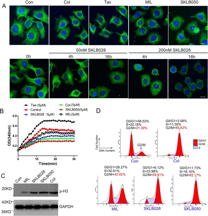
Figure 2.
MDs inhibit tubulin polymerization and cause G2/M phase cell-cycle arrest. A, HCT-8/V cells were treated with 1 μm MIL, 50 nm SKLB050, 50 nm colchicine, or 50 nm paclitaxel for 16 h or treated with 50 nm or 200 nm SKLB028 for 4 or 16 h. Then tubulin morphology was detected by immunofluorescence with anti-β-tubulin and DAPI staining. Bar, 10 μm. B, indicated concentrations of compounds were co-incubated with tubulin (3 mg/ml) at 37 °C. Absorbance at 340 nm was detected every 1 min for 30 min. C, HCT-8/V cells were incubated with 1 μm MIL, 50 nm SKLB050, 50 nm colchicine, or 100 nm SKLB028 for 16 h; then cell lysates were collected and subjected to Western blotting for p-H3 detection. GAPDH was used as a loading control. D, HCT-8/V cells were incubated with 1 μm MIL, 50 nm SKLB050, 50 nm colchicine, and 100 nm for 16 h; subjected to PI staining; and analyzed by a flow cytometer for cell-cycle analysis. Col, colchicine; Con, control; Tax, paclitaxel.

