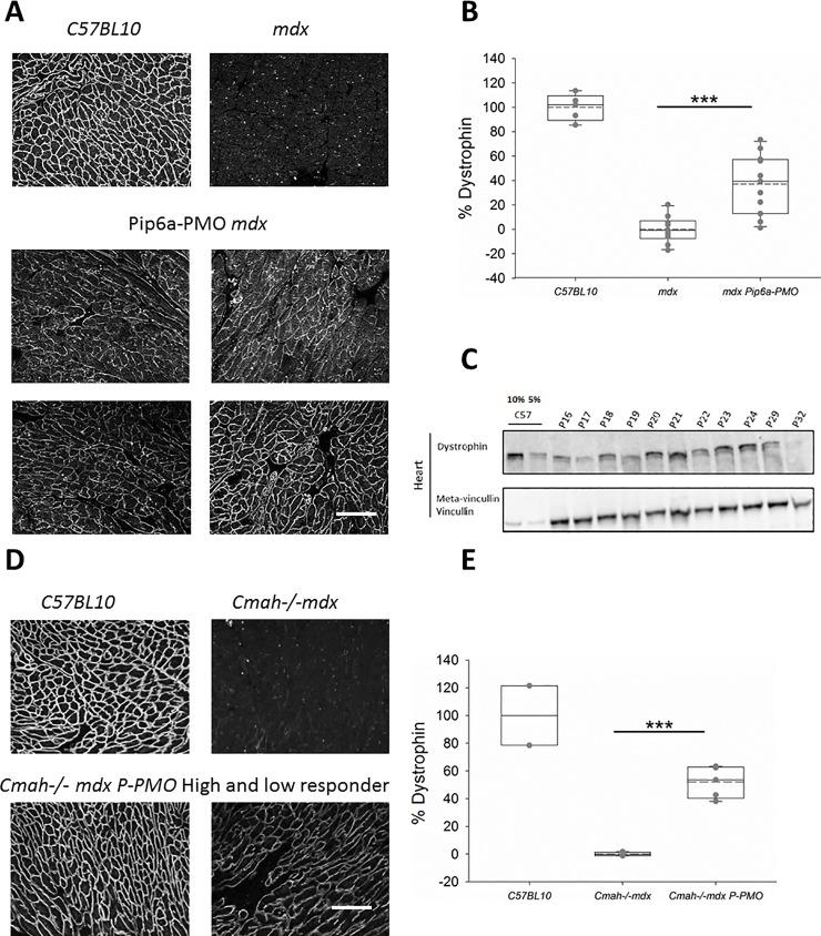
Fig 1.
A-E: Pip6a-PMO restores cardiac dystrophin in mdx and Cmah-/-mdx mice. A) Immunofluorescent (IF) staining of dystrophin in C57BL10, control mdx and four P-PMO treated mdx mice demonstrating the homogeneity and range of restoration. B) Box plots showing quantification of dystrophin restoration on dystrophin/laminin IF. Average dystrophin restoration was 37.2% (***p<0.001, Unpaired Student’s t-test) (C57BL10 n = 6, mdx n = 8 and P-PMO treated mdx n = 11 mice). C) western blot of cardiac dystrophin expression and meta-vinculin control in individual P-PMO treated mdx mice and C57BL10 mice (10% and 5%), again demonstrating the range of restoration. mdx control samples which showed no band for dystrophin were ran in parallel on a separate gel. D) IF staining of dystrophin in C57BL10, control Cmah-/-mdx and examples of a high and low ‘responder’ (mice showing the best and worst levels of dystrophin restoration, respectively). E) Box plots showing quantification of dystrophin restoration on dystrophin/laminin IF. Average dystrophin restoration was 51.6%. (***p<0.001, Unpaired Student’s t-test). Scale bars = 100 μm (C57BL10 n = 2, Cmah-/-mdx n = 6 and P-PMO treated Cmah-/-mdx 6 mice/group).

