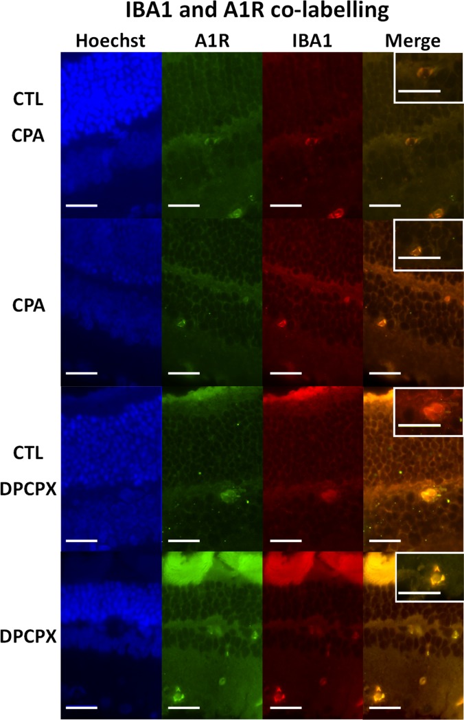
Fig 3. Double immunolabeling for Iba 1 and A1 receptor.
Representative sections of CPA Control retina (top row); CPA treated retina (second row); DPCPX Control retina (third row) and DPCPX treated retina (fourth row). In every case nuclear staining with Hoechst 33258 (blue), A1 receptor immunolabeling (green); Iba 1 immunolabeling (red), and double labeling of the same sections may be observed form left to right. Insets show higher magnifications of merge images. Scale bars = 20 μm.

