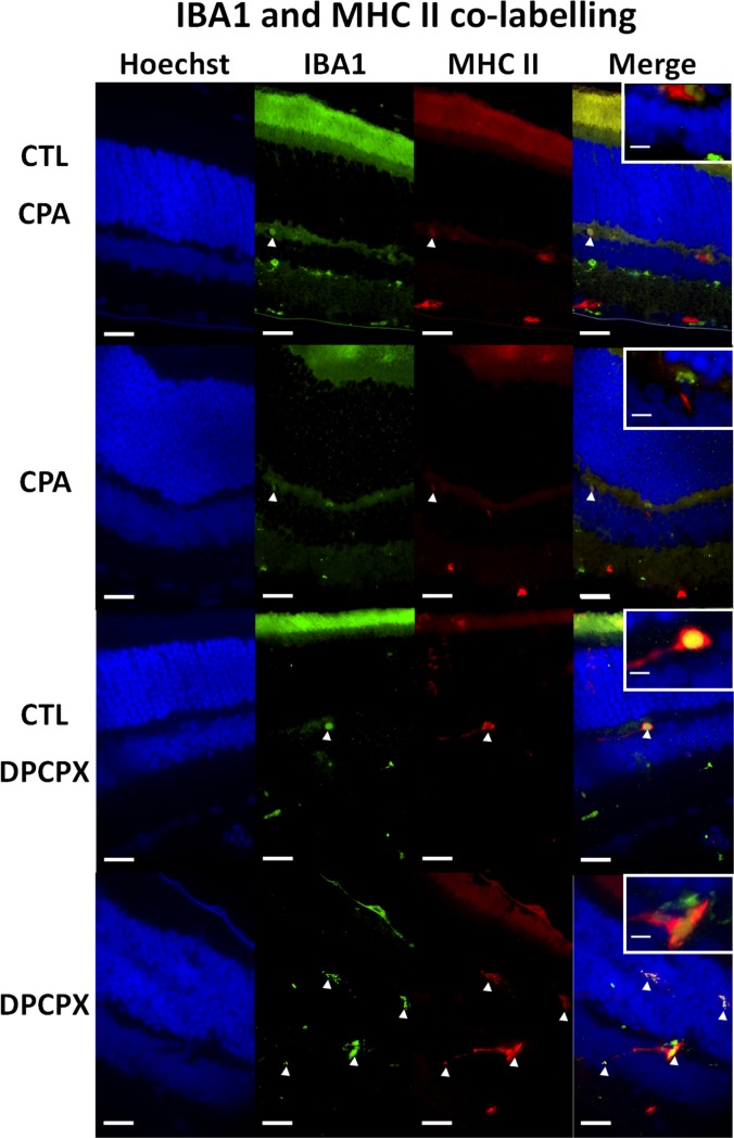
Fig 4. Double immunolabeling for Iba 1 and MHC-II.
Representative sections of CPA Control retina (top row); CPA treated retina (second row); DPCPX Control retina (third row) and DPCPX treated retina (fourth row). In every case nuclear staining with Hoechst 33258 (blue), Iba 1 immunolabeling (green), MHC II immunostaining (red) and double labeling (merge) of the same sections may be observed form left to right. Insets show higher magnifications of merge images. Arrow heads show reactive microglial cells. Observe the low number of double stained reactive microglial cells in CPA treated retina and the higher number of double stained reactive microglial cells in DPCPX treated retina. Scale bars = 20 μm and 5μm (inset).

