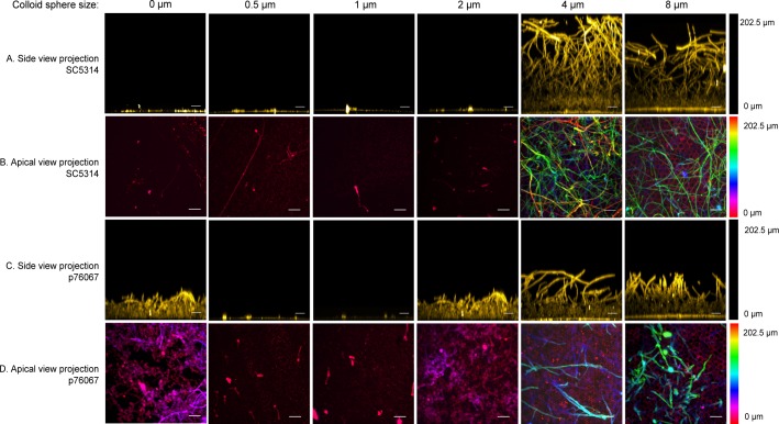
Fig 4. Confocal images of biofilm growth on colloid crystal surfaces.
Wild-type C. albicans strains SC5314 (rows A, B) and p76067 (rows C, D) were grown on test solids with colloid crystal sphere diameters indicated above each column, then fixed and stained with ConA Alexa Fluor 594 as detailed in Methods. Rows A and C show side view projections; rows B and D show apical projections. The scale bar corresponds to 20μm. The hyphae are the long fibrous features; the yeast cells the ~3μm oval features. The 0 μm sample refers to PDMS with a layer of silica grown on it. This solid has nanometer-scale roughness but no micrometer-scale features. In this sense, it is equivalent to a coating of zero-μm spheres.

