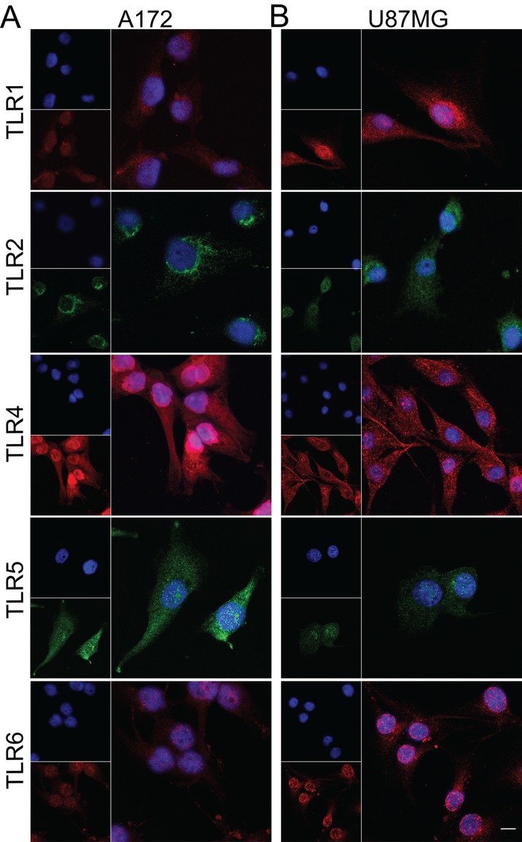
Fig 3. Immunofluorescence of TLR1, TLR2, TLR4, TLR5, and TLR6 in GBM cell lines.
A172 (A) and U87MG (B). TLR1, TLR4, and TLR6 are stained in red, TLR2 and TLR5 in green, and nuclei in blue by DAPI. The presence of all five TLRs was detected in both cell lines. Expression of TLR5 was more intense in A172 compared to U87MG. TLR4 and TLR5 positivity were detected in both tumor lineage cells nuclei. Magnification of 400x.

