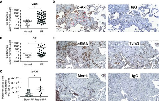
Figure 1.
Gas6 and Axl expression and activation in idiopathic pulmonary fibrosis (IPF). (A and B) Gas6 (A) and Axl (B) transcript expression were analyzed in surgical lung biopsies from nondiseased patients (normal; n = 10) and patients with IPF (n = 42) by quantitative PCR. Patients with IPF were stratified as rapid or slow progressors as defined previously (19). (C) p-Axl expression was quantified via software analysis (see Figure E1) in biopsies from patients with slowly and rapidly progressing IPF. (D and E) Representative p-Axl, α-smooth muscle actin, Tyro3, and Mertk in contiguous IPF lung sections. IgG control staining for the same areas is shown in both panels. Data in A and B are mean ± SD and in C mean ± SEM. Scale bars, 100 μm. *P ≤ 0.05. Red arrows highlight fibroblasts stained for pAxl. α-SMA = α-smooth muscle actin; Axl = anexelekto; Gas6 = growth arrest–specific 6; Mertk = MER proto-oncogene, tyrosine kinase; p-Axl = phosphorylated Axl; Tyro3 = TYRO3 protein tyrosine kinase 3.

