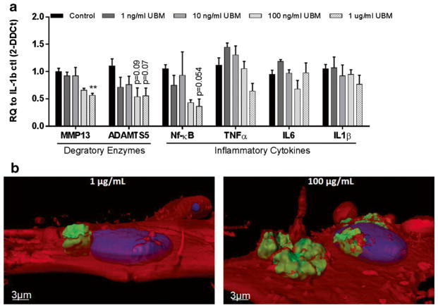
Fig. 6.
UBM decreased inflammatory markers in human primary chondrocytes a Gene expression data from in vitro human chondrocytes exposed to 10 ng/mL of IL-1β and 1 ng/mL–1 μg/mL of UBM (n = 3). *p < 0.05, **p < 0.01. b Confocal imaging of 1 μg/mL UBM (left) and 100 μg/mL UBM (right) 24 h after addition into cell culture medium. Red = cell membrane (seen at 50% transparency), blue = nucleus, green = UBM. UBM particles appear to be almost entirely encapsulated by the cell membrane after only 24 h

