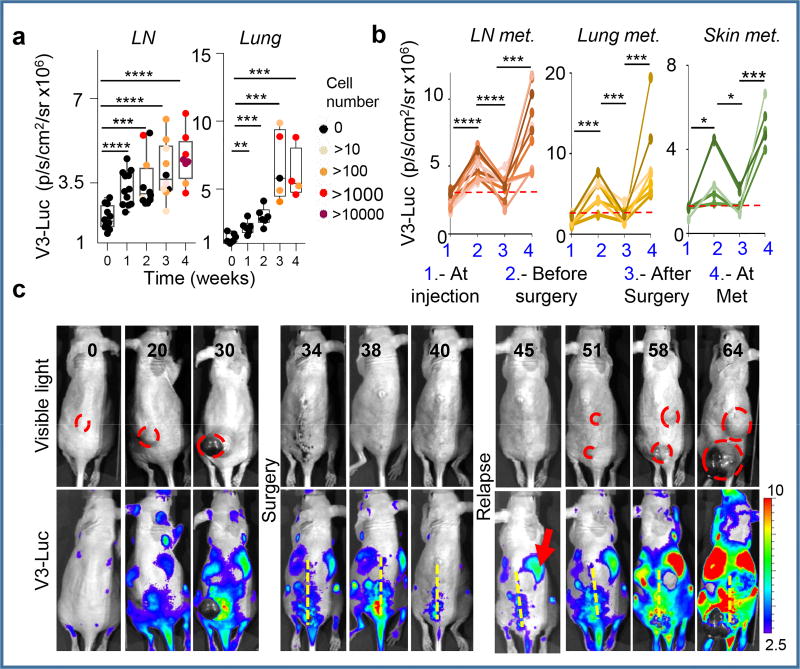
Figure 2. Vegfr3Luc mice reveal pre-metastatic niches.
a, V3-Luc emission by xenografts of mCherry-SK-Mel-147 in sentinel LN and lungs. Colored dots correspond to tumor cell burden defined by RT-PCT. b, Quantification of V3-Luc emission by SK-Mel-147-mCherry prior and after surgical resection of the cutaneous lesions. LN (n=9), lung metastases (n=7) and skin metastases (n=5). t-Test. c, representative whole-body imaging of experiments as in (b). See Source Data for V3-Luc quantifications in panels (a,b).

