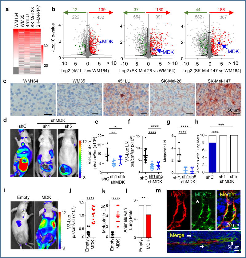
Figure 3. Proteomic analyses identify MIDKINE as new pro-lymphangiogenic/pro-metastatic factor.
a, LFQ (Label Free Quantitative) expression of exosome cargo identified by LC/MS-MS in the indicated cell lines. b, Volcano plots showing exosomal proteins (grey dots) differentially expressed with respect to the non-lymphangiogenic WM164. Proteins upregulated or downregulated vs. the poorly-metastatic WM35 are depicted in red or green, respectively. The hyperbolic black curve separates statistically-significant regulated proteins as defined by two-sample Student’s t-Test, FDR=0.05; S0=0.8. c, Immunological detection of MDK (pink) in tumor xenografts. d, V3-Luc emission by SK-Mel-147-mCherry transduced with control or MDK shRNA(1),(5), with quantifications for cutaneous lesions in (e) and for sentinel and brachial LN in (f). g, Metastatic LN (mCherry positive) of animals in (e). Data correspond to mean ± SD (n≥ 6 mice/condition). One-way ANOVA/Dunnett's correction for multiple comparison. h, Lung metastases (blue) after tumor removal. Fisher's exact test, n=10. i, V3-Luc emission by MDK overexpression in the otherwise negative WM164. Scale, p/s/cm2/sr (×106). j, Relative nodal V3-Luc emission at the indicated conditions. The number of metastatic LN per animal is shown in panel (k). t-Test. i, Lung metastases (mCherry positive; red) in MDK GoF studies (generated by overexpressing MDK in the negative WM164). Fisher's exact test, n=7. m, Immunomicrographs showing MDK (green) accumulated at areas of neo-lymphangiogenesis and at sites of lymphatic sprouting in LN (arrows). Asterisks mark stromal cells. See Source Data for V3-Luc quantifications of panels (e–h, j–l) and Supplemental Figure 1 for uncropped blots.

