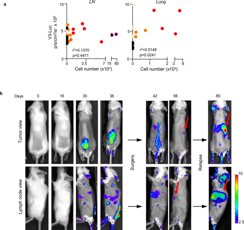
Extended Data Figure 4. Analysis of metastatic potential in Vegfr3Luc mice.
a, Pearson correlation analyses of the luciferase signal vs cell burden, corresponding to data presented in Fig. 2a. Shown are lymph nodes or lungs with luciferase signal over the background. b, V3-Luc emission in immunocompetent BrafV600E;Ptenlox/lox (albino) mice at the indicated times prior and after surgical removal of the primary cutaneous melanoma. Note the reduction of tumor-driven Vegfr3-Luc signal particularly in visceral sites, and the reactivation at later time points (black arrows), marking metastatic relapse. Fur was removed to ease in the imaging.

