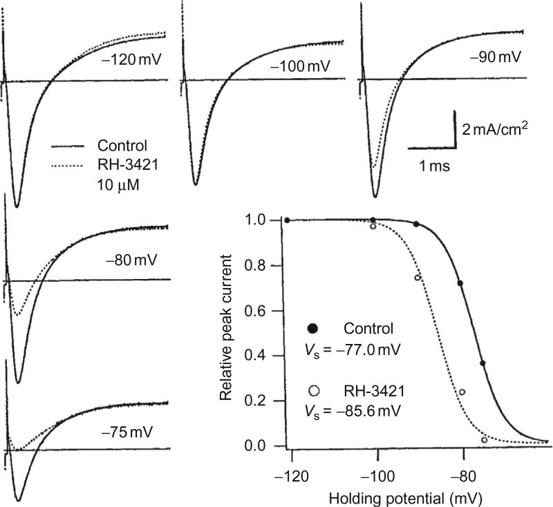
Figure 5.7.
Dihydropyrazole block appears as a parallel shift of the steady-state slow inactivation curve in the direction of hyperpolarization. Ionic current traces from voltage-clamped crayfish giant axons were scaled by a common factor so that the peak at −120 mV matched the peak before treatment with the dihydropyrazole. Peak/Na was depressed most at depolarized potentials, whereas outward current, /K, was not affected by the treatment. The graph shows plots of peak current normalized to the value at − 120mV. RH-3421 (10µM) appears to shift the steady-state inactivation relation to the left by 8.6 mV. Reproduced from Salgado (1992) with permission from the American Society for Pharmacology and Experimental Therapeutics.

