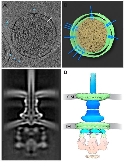
Figure 3.
The Type III Secretion System in situ. (A) Slice through a tomogram of a Salmonella Typhimurium minicell, showing multiple secretion systems (blue arrows) crossing the inner (IM) and outer (OM) membranes. (B) Segmentation of the tomogram in (A). (C) Cross-section through 3D average of the T3SS, showing the outer membrane (OM) and inner membrane (IM), as well as the cytoplasmic complex and needle portions of the T3SS. From [32]. (D) 3D cryo-EM reconstruction of the T3SS shown in (C) (EMD 8544).

