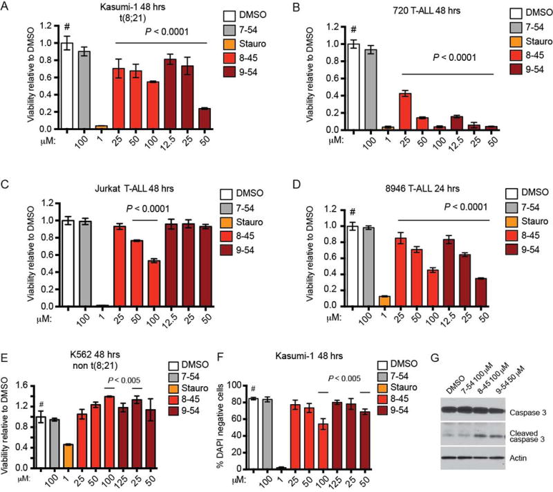
Figure 4. RDIs reduce cell growth and induce apoptosis in leukemia cells.

A–E. RDIs AI-8–45 (8–45) and AI-9–54 (9–54) inhibit proliferation of the AML cell line Kasumi-1 and the T-ALL lines 720, Jurkat (8–45 only), and 8946, but not K562 as detected by MTT cell viability assay. AI-7–54 (7–54) is the negative control, and staurosporine (Stauro) is a positive control. Data represent mean values for triplicates ± standard deviation (SD) (two independent experiments). P values were calculated by one-way ANOVA (staurosporine-treated cells were not included in the ANOVA analysis). Dunnett’s Multiple Comparison test was performed using DMSO treated cells as the comparator (#); horizontal lines above columns indicate significant differences from DMSO treated cells (P ≤ 0.05).
F. RDIs reduce the percentage of live (DAPI negative) Kasumi-1 cells as measured by flow cytometry. Data represents mean values of two independent experiments; statistical analysis as in A–E.
G. RDI treatment results in increased caspase-3 cleavage in 720 T-ALL cells (48 hrs).
