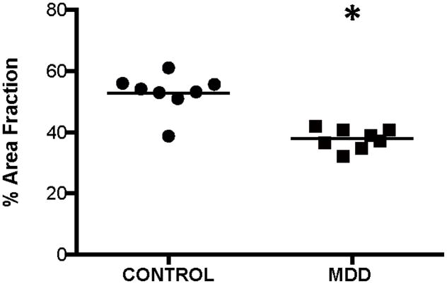
Figure 4.
Area fraction of all GFAP-immunoreactive structures (cell bodies + processes) in the ventral prefrontal white matter of human brain. Area fraction was significantly decreased (ANCOVA, F(1,10)=43.322, *p<0.0001) in subjects with major depressive disorder (MDD) as compared to control subjects. The horizontal lines represent the mean value of density for each diagnostic group.

