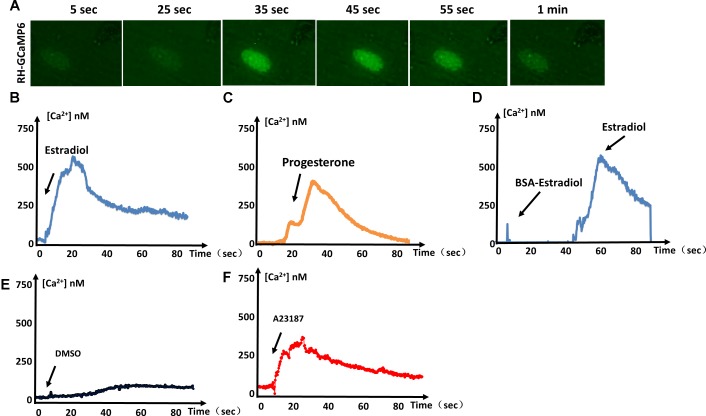
FIGURE 1.
Estrogen-induced Ca2+ fluxes in T. gondii. (A) Video microscopy of intracellular parasites expressing GCaMP6f, following the addition of estradiol at 0 s. (B) Intracellular Ca2+ concentrations monitored over time in GCaMP6f-RH parasites (extracellular tachyzoites) stimulated with 10-8 M estradiol. (C) Intracellular Ca2+ fluxes of GCaMP6f-RH parasites were monitored after stimulation with 10-5 M progesterone. (D) GCaMP6f-RH parasites stimulated with estradiol after treatment with BSA-coupled estradiol. (E) DMSO was used as a negative control because all compounds dissolved in DMSO. (F) A23187 (calcium ionophore, 2 μM, Sigma, United States), a mobile ion-carrier, was used as a positive control for the monitoring of intracellular Ca2+ concentrations. Each experiment was performed in triplicate with three independent biological replicates.

