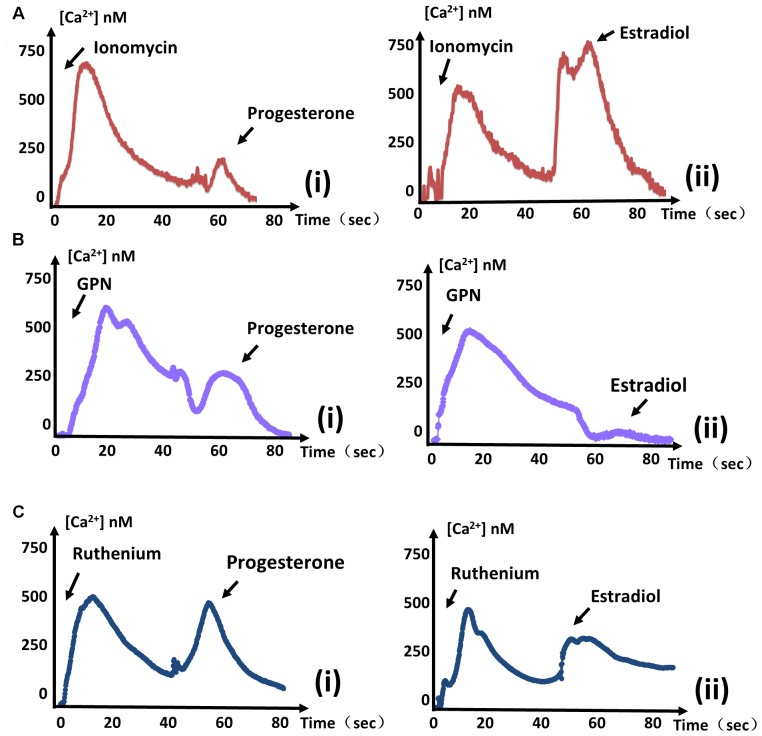
FIGURE 2.
Estradiol-induced Ca2+ and progesterone-induced Ca2+ originate from different stores. (A) The same measurements were performed as in Figure 1. Ionomycin was added at 0 s, and progesterone (i) or estradiol (ii) was added at 60 s to monitor intracellular Ca2+ concentrations. (B) The intracellular calcium concentrations of parasites were measured after treatment with GPN at 0 s. Progesterone (i) or estradiol (ii) was added at 60 s, as indicated. (C) GCaMP6f-RH parasites were treated with ruthenium red at 0 s, and stimulated with progesterone (i) or estradiol (ii) at 40 s to observe Ca2+ fluxes. Each experiment was performed in triplicate with three independent biological replicates.

