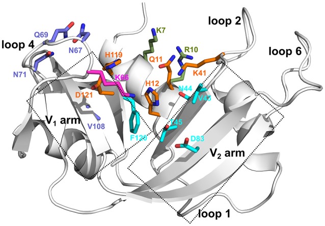
Figure 1.

Ligand binding subsites identified in bovine RNase A. Residues of the ligand binding subsites, corresponding to the base B1, B2, and phosphate P0, P1, and P2 subsites are represented as sticks colored cyan, purple, magenta, orange, and olive, respectively. Catalytic residues His12, Lys41, and His119 form part of the P1 binding site.
