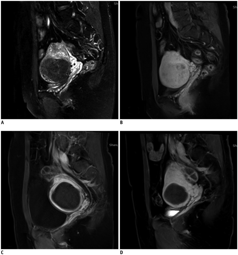
Fig. 3. Patient with symptomatic uterine fibroids treated by USgHIFU.
Representative images are shown for three different time points. A. Uterine fibroid in 36-year-old woman was hypo-intense on pretreatment T2WI. B. Sagittal contrast-enhanced T1WI showed homogeneous enhancement before USgHIFU. C. Sagittal contrast-enhanced T1WI showed 100% NPV in fibroid immediately after treatment. D. Fibroid volume shrinkage (64.5% of baseline) with sustained non-perfused area at 6-month follow-up.

