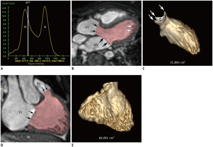
Fig. 2. 10-year-old female with repaired tetralogy of Fallot.
A. Histogram shows threshold of 497 (white line) located between two different distribution curves of M and ventricular B. Threshold is close to myocardium curve to exclude tissues consisting of 100% ventricular myocardium and it was used for 3D threshold-based segmentation method. B. LV long-axis reformatted image using ES 3D whole-heart MRI demonstrates segmented LV cavity in pink. Even after exclusion of papillary muscles and trabeculations from ventricular cavity by using 3D threshold-based segmentation, small fraction of pixels partially including myocardial tissue are present along LV endocardium (white arrows). AV and MV were manually segmented (black arrows). C. LV ESV segmented by using 3D threshold-based method and 3D whole-heart MRI data was 31.8 mL. Three commissures (arrows) of aortic valve are clearly noted. D. RV long-axis reformatted image using ES 3D whole-heart MRI displays segmented RV cavity in pink. Although papillary muscles and trabeculations are excluded from ventricular cavity by using 3D threshold-based segmentation, small fraction of pixels partially including myocardial tissue are present along RV endocardium. PV and TV were manually segmented (arrows). E. RV ESV segmented by using 3D threshold-based method and 3D whole-heart MRI data was 66.1 mL. AV = aortic valve, B = blood, ESV = end-systolic volume, M = myocardium, MV = mitral valve, PV = pulmonary valve, TV = tricuspid valve

