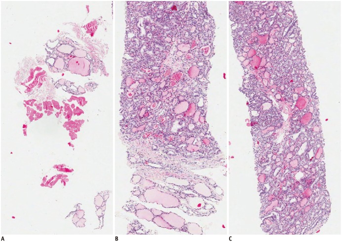
Fig. 1. Core biopsy specimens using modified CNB technique, H&E staining (× 40).
This case was diagnosed as follicular variant of papillary thyroid carcinoma.
Biopsy specimens were retrieved from surrounding normal parenchyma (A), capsular portion of nodule (B), and intra-nodular target (C). CNB = core needle biopsy, H&E = hematoxylin and eosin

