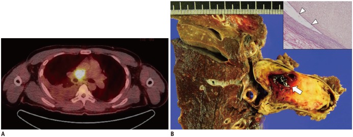
Fig. 4. FDG positron emission tomography-CT image obtained from 24-year-old patient with PAIS (same patient in Fig. 1C).
Surgical resection was performed.
A. Axial image shows high FDG uptake in right pulmonary artery (maxSUV: 12.9). B. Focal hemorrhage in mass of gross specimen (arrow); this is common in PAIS (arrow). High magnification elastic Van-Gieson-stained specimen (inset, × 100) shows mass of intimal origin detached from intimal layer (arrowheads). FDG = fluorodeoxyglucose

