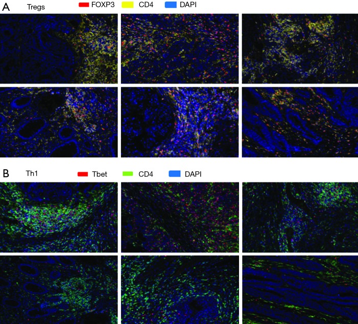
Figure 4.
Multispectral fluorescent IHC staining (200×) and image analysis using a panel of markers including CD4, FOXP3, and Tbet on pretreatment tumor specimens from 6 patients with MSI-H or POLE-mutated metastatic colorectal tumors identified (A) expression of CD4 and FOXP3 within the TME of responders (top row) and nonresponders (second row from top) to PD-1/PD-L1/CTLA-4 blockade, while (B) a greater amount of CD4+ Tbet+ cells was identified within the TME of responders (third row from top) compared to nonresponders (bottom row). Cases stained for FOXP3 and CD4, clockwise fashion starting from top left panel, in (A) from a MSI-H mCRC responder to pembrolizumab, MSI-H mCRC responder to pembrolizumab, MSI-H mCRC responder to investigational anti-PD-L1 agent and tremelimumab, MSI-H mCRC nonresponder to pembrolizumab, MSI-H mCRC nonresponder to pembrolizumab, and POLE-mutated mCRC nonresponder to pembrolizumab. Cases in (B) correspond to cases in (A) in same order but stained for Tbet and CD4. Multispectral fluorescent IHC stains: (A) FOXP3 (red), CD4 (yellow), DAPI (blue); (B) Tbet (red), CD4 (green), DAPI (blue). IHC, immunohistochemistry; PD-1, programmed cell death 1; PD-L1, programmed death ligand 1; DAPI, 4',6-diamidino-2-phenylindole; MSI-H, microsatellite instability-high; mCRC, metastatic colorectal cancer; TME, tumor microenvironment; CTLA-4, cytotoxic T lymphocyte-associated protein 4.

