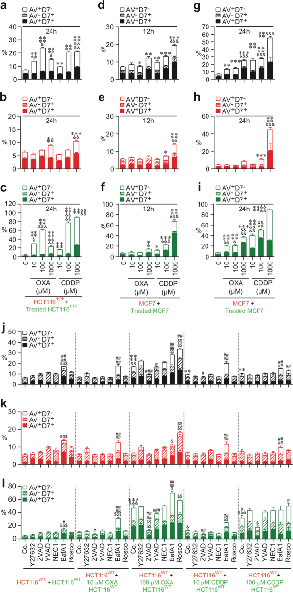
Fig. 4. Quantitative imaging flow-cytometric detection of CAD modalities induced by oxaliplatin or cisplatin.
The cell death profiling was performed after co-cultures of untreated (red) CMTMR-labeled HCT116WT, HCT116+/+, or MCF7 cells with untreated (green) CMFDA-labeled cells or with oxaliplatin (OXA)-treated or cisplatin (CDDP)-treated (green) CMFDA-labeled HCT116WT, HCT116+/+, or MCF7 cells. Co-cultures have been performed during 12 h (d–f) and 24 h (a–c, g–l) with the indicated concentrations of OXA or CDDP in the presence or absence of death effector inhibitors. As previously described, cells were labeled for the simultaneous detection of type I, II, or III cell death. Means ± SEM are indicated (n = 3). For a–c, asterisk (*) is used for the comparison of “HCT116+/++OXA- or CDDP-treated HCT116+/+” with “HCT116+/++0 nM HCT116+/+” for AV+D7−, hash (&) is used for the comparison of “HCT116+/++OXA- or CDDP-treated HCT116+/+” with “HCT116+/++0 nM HCT116+/+” for D7+. For d–i, asterisk (*) is used for the comparison of “MCF7+OXA- or CDDP-treated MCF7” with “MCF7+0 nM MCF7” cells for AV+D7− and ampersand (&) is used for the comparison of “MCF7+OXA- or CDDP-treated MCF7” with “MCF7+0 nM MCF7” for D7+. For j–l, asterisk (*) is used for comparison of “HCT116WT+OXA- or CDDP-treated control (Co.) HCT116WT” with “HCT116WT+control (Co.) HCT116WT” for AV+D7−, ampersand (&) for the comparison of “HCT116WT+OXA- or CDDP-treated control (Co.) HCT116WT” with “HCT116WT+control (Co.) HCT116WT” for D7+, hash (#) for the comparison of inhibitor-treated cells with respective control cells for AV+D7−, and dollar symbol ($) for the comparison of inhibitor-treated cells with respective control cells for D7+. *, #, $, &p < 0.05; **, ##, $$, &&p < 0.01; ***, ###, $$$, &&&p < 0.001; and ****, ####, $$$$, &&&&p < 0.0001

