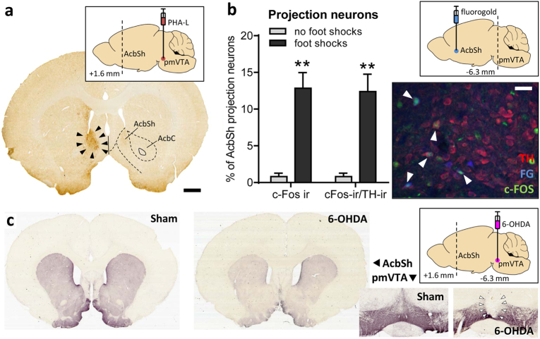
Fig. 1.
The pmVTA projection to the AcbSh was activated by exposure to foot shocks. a Triangles indicate terminals in AcbSh arising from the pmVTA, as labeled by an iontophoretic injection with the anterograde tracer PHA-L into the pmVTA (inlay; see also Supplementary Figure 1). Scale bar: 1 mm. b FG was injected into the AcbSh (inlay; see also Supplementary Figure 2). Then one group of rats were exposed to foot shocks (n = 4) whereas another group received no shocks (n = 5). The photomicrograph shows a representative section of the pmVTA with TH-, FG-, and c-Fos-positive neurons. White triangles indicate triple-labeled neurons. The bar diagram depicts the percentage of pmVTA-AcbSh projection neurons (i.e. fluorogold-positive) which is also c-Fos immunoreactive (ir) or c-Fos and TH-ir. Scale bar: 100 µm. **p < 0.01, Student's t-test, comparison with the no-shocked group. c In experiment 2, saline (n = 9) or 6-OHDA (n = 23) was injected into the pmVTA (inlay). The photomicrographs are from representative TH-immunostained coronal sections. 6-OHDA injections strongly reduced TH-immunoreactivity within the pmVTA (right panel) and the AcbSh (left panel). Based on histological analyses, rats were divided into groups with misplaced lesions (n = 7) and lesions restricted to the pmVTA (here indicated by triangels; n = 16). For behavioral data, see Fig. 2

