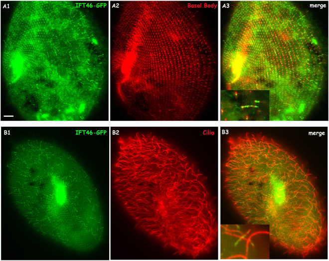
Figure 5.
Co-localisation of the IFT46-GFP fusion protein on basal bodies and cilia is displayed by using ID5 and anti-polyglycylated tubulin antibodies, respectively, as markers. (A1,B1) Green fluorescence displays the localisation of the IFT-46 fusion protein. (A2) Basal bodies stained by the ID5 antibody against polyglutamylated tubulin. (B2) Cilia labelled by anti-polyglycylated tubulin antibody. (A3,B3) Merged photos show co-localisation of the IFT46-GFP fusion protein on basal bodies and cilia, and magnifications show details in cilia and basal bodies. Bar: 10 µm.

