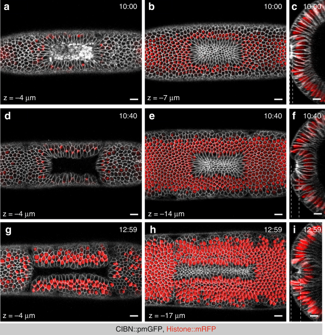
Fig. 5.
Sustained RhoGEF2 optogenetic activation is sufficient to drive a complete invagination. a–i Confocal images of three individual embryos co-expressing CIBN::pmGFP, RhoGEF2-CRY2, and Hist2A::mRFP as nuclear marker. Within a rectangular on the dorsal epithelium, two-photon illumination was restricted to three consecutive z-stacks (1 µm spacing) centered at 4 µm from the apical-most plane, and excited for ~152 ms during each acquisition round. Sustained photo-activation was alternated with mCherry excitation at intervals of ~13 s. As the epithelium folded inwards, continuous two-photo-activation was achieved by manually adjusting the ROI such that illumination was restricted to the initial group of photo-activated cells. In addition, the focal plane of activation was manually rectified to match the cells’ apical-most plane. At the indicated time points, a z-stack comprising the dorsal epithelium was acquired for both mRFP and GFP (50 planes, 1 µm spacing). Overlays of both channels are shown. a–c Three views of the dorsal epithelium after 10:00 min of continuous activation. a Surface view of the dorsal epithelium at a depth of 4 µm. b Apices of activated cells at a depth of 8 µm. c Cross-section view of the activated area. d–f Three views of the dorsal epithelium after 10:48 min of continuous activation. d Surface view of the dorsal epithelium at a depth of 4 µm. e Apices of activated cells at a depth of 14 µm. f Cross-section view of the activated area. g–i Three views of the dorsal epithelium after 12:59 min of continuous activation. g Surface view of the dorsal epithelium at a depth of 4 µm. h Apices of activated cells at a depth of 17 µm. i Cross-section view of the activated area. Dashed lines indicate the depths at which surface views were acquired (left and middle panels). Scale bars, 10 µm

