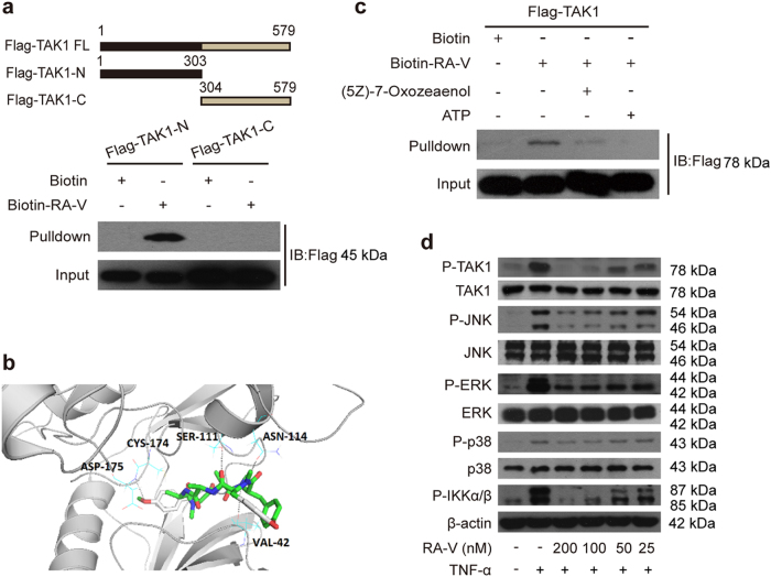
Fig. 6. RA-V binds the kinase domain of TAK1.
a CB12 directly binds to the TAK1 kinase domain. HEK293T cells were transfected with TAK1-N or TAK1-C for 24 h. The cells lysates were incubated with CB12 or biotin and precipitated with streptavidin-coated sepharose and were analyzed by immunoblotting with Flag antibody. b The model for RA-V binding to the crystal structure of the TAK1–TAB1 fusion protein (PDB 4GS6). The blue dotted lines represent the hydrogen bonds between RA-V (green) and TAK1–TAB1. c RA-V probably binds the ATP-binding pocket of TAK1. HEK293T cells were transfected with TAK1 for 24 h, and then the cell lysates were incubated with biotin or CB12 before precipitation with streptavidin agarose. The immunoprecipitates were treated with ATP or (5Z)-7-oxozeaenol and western blotted with Flag antibody. d RA-V inhibits TAK1, JNK, ERK, p38, and IKKα/β phosphorylation. HeLa cells were incubated with various concentrations of RA-V for 12 h and then treated with 10 ng/mL TNF-α for 10 min. The cell lysates were prepared and western blotted with the indicated antibodies. Each experiment was repeated at least three times

