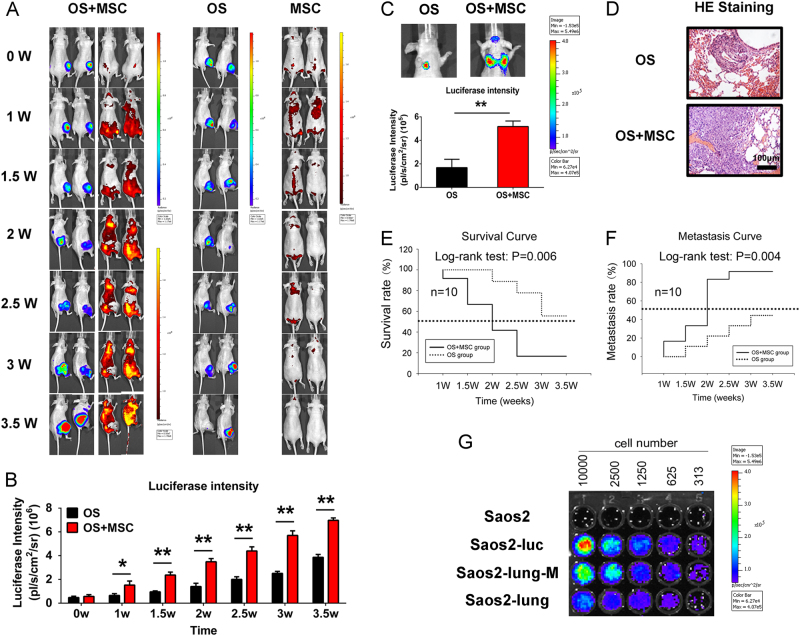
Fig. 2. MSCs enhanced OS metastasis in OS-bearing nude mice.
a The interactions between MSCs and OS cells were monitored in vivo using the IVIS 200 system. b Quantification of luciferase intensity. Data are presented as mean ± standard deviation (S.D.). *p < 0.05 and **p < 0.01. Each group contained 10 animals. c Pulmonary metastasis of OS was facilitated by interactions with MSCs. d HE staining of pulmonary metastasis of OS. e Survival–time curve indicating that MSCs shortened the survival of OS-bearing mice. f Metastasis–time curve indicating that MSCs promoted the pulmonary metastasis of OS. g Identification of isolated luciferase-labeled OS cells that underwent pulmonary metastasis in the presence or absence of the MSC-CM stimulus. Data are presented as mean ± S.D. *p < 0.05, **p < 0.01. All in vitro data were obtained from at least three independent experiments. Each group contained 10 animals

