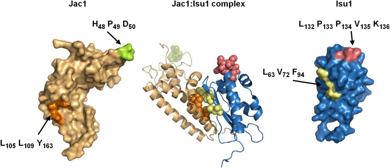
Fig. 3.
Model of the Jac1–Isu1 complex (center panel) on the basis of in silico docking of the Jac1 protein crystal structure (PDB code 3UO3, left panel) and homology model of the Isu1 structure (right panel). Residues of Jac1 and Isu1 implicated in their interaction are highlighted [51]

